


Coxarthrosis- This is arthrosis of the hip joint.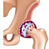 It develops gradually, for several years, predisposed to progression, can be both double.It is accompanied by pain and restriction of movement in the joint.In the short stages there are hip muscle atrophy and shortening of the limb.The diagnosis is established on the basis of clinical symptoms and radiography results.In the early stages of coxarthrosis, conservative treatment.With the destruction of the joint, especially in patients of young and middle age, surgery (endoprothetics) has been indicated.
General information
Coxarthrosis (osteoarthrosis or deformation of arthrosis of the hip joint) is a degenerative-dystrophic disease.It usually develops at the age of 40 and more.This can be the result of various injuries and joint diseases.Sometimes it happens for no apparent reason.Coxarthrosis is characterized by a gradual progressive course.Conservative treatments are used in the early stages.In the short stages, the joint function can only be restored operational.
Coxarthrosis is one of the most common arthrosis in orthopedics and traumatology.The high frequency of its development is due to a significant stress of the hip joint and the widespread spread of congenital pathology - joint dysplasia.Women suffer from coxarthrosis a little more often than men.
The causes of coxarthrosis
The main (occurring for unknown reasons) and secondary (developed as a result of other diseases) arthrosis of the hip joint are distinguished.
Secondary coxarthrosis can be the result of the following diseases:
- Hip dysplasia.
- Congenital dislocation of the thigh.
- Pertes diseases.
- Aseptic necrosis of the thigh head.
- Infectious lesions and inflammatory processes (such as hip arthritis).
- Injuries (traumatic dislocations, fractures of the hip joint, pelvic fractures).
Coxarthrosis can be either one or double.With primary coxarthrosis, a concomitant lesion of the spine (osteochondrosis) and the knee joint (gonarthrosis) is often observed.
Risk factors
Among the factors that increase the likelihood of developing coxarthrosis include:
- Constant increased joint load.Most often, they are observed in athletes in people with excess weight.
- Circulatory disorders, hormonal changes, metabolic disorders.
- Spinal pathology (kyphosis, scoliosis) or stopping (flat legs).
- Adult and senile age.
- A sedentary lifestyle.
Coxarthrosis itself is not inherited.However, some characteristics (metabolic disorders, structural characteristics of the skeleton and the weakness of the cartilage) may be inherited by the child by the parents.Therefore, in the presence of blood relatives suffering from coxarthrosis, the likelihood of the disease is slightly increased.
Patanatomy
The hip joint is formed by two bones: ileum and femur.The hip head is articulated with the acetabulum of the iliac bone, forming a special "hinge".During the movements, the acetabulum remains stationary, and the head of the femur moves in different directions, providing flexion, expansion, abduction, wearing and rotating hips.
During the movements, the joint surfaces of the bones are unobstructed slides to each other, thanks to the smooth, elastic and durable cartilage of hyaline, covering the cavity of the rotating cavity and the head of the thigh.In addition, Hyaline cartilage performs a shock aspire function and participates in the redistribution of load while moving and walking.
There is a small amount of joint fluid in the joint cavity that plays the role of lubrication and provides a feeding of hyaline cartilage.The joint is surrounded by a thick and strong capsule.Above the capsule are large hip and gluteal muscles, which provide movements in the joint and together with hyaline cartilage are also shock absorbers that protect the joint from injury with unsuccessful movements.
With coxarthrosis, the joint fluid becomes greater and more viscous.The surface of the hyaline cartilage dries, loses smoothness covered with cracks.Due to the roughness, cartilage during movements is constantly wounded for each other, which causes them to thin and impair pathological changes in the joint.As coxarthrosis progresses, bones begin to deform, "adapting" to increased pressure.Metabolism in the joint worsens.In the short stages of coxarthrosis there is severe atrophy of the muscles of the inflamed limb.
Symptoms of coxarthrosis
The main symptoms of the disease include pain in the joint, inguinal region, thigh and knee joint.Coxrtrosis, stiffness and stiffness of the joint are also observed, gait disruption, lame, hip atrophy and limb contraction on the side of the lesion.A characteristic characteristic of coxarthrosis is a limitation of abduction (for example, the patient is difficult when he tries to sit in a chair).The presence of certain signs and their severity depends on the stage of coxarthrosis.The first and most constant symptom is pain.
AtFirst -degree coxarthrosisPatients complain of periodic pain that occurs after physical activity (running or long walking).The pain is localized in the joint, rarely in the thigh or knee.After rest, it usually disappears.The first -degree coxarthrosis gait is not disturbed, the movements are fully preserved, there is no muscular atrophy.
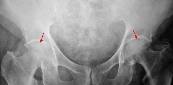
The X -ray patient suffering from first -degree coxarthrosis is determined by mild changes: moderately uneven narrowing of the joint, as well as bone plants around the outer or inner edge of the acetabulum in the absence of changes from the head and neck of the femur.
AtCoxarthrosis 2 degreesThe pain becomes more intensive, often appearing at rest, radiating in the thigh and groin.After considerable physical activity, the patient with coxarthrosis begins to limp.The volume of movements in the joint decreases: the abduction and internal rotation of the thigh is limited.
In X -Remitt images for coxarthrosis of the 2nd degree, a significantly uneven narrowing of the joint gap (more than half of the normal height) is determined.The head of the femur is somewhat displaced up, deformed and increases in size, and its contours become uneven.Bone growths with this degree of coxarthrosis appear not only on the internal but also on the outer edge of the acetabulum and go beyond the cartilage.
AtCoxarthrosis 3 degreesThe pain becomes constant, concern for patients not only during the day but also at night.Walking is difficult when moving, a patient with coxarthrosis is forced to use a cane.The volume of movements in the joint is sharply restricted, the muscles of the ass, hips and lower legs are atrophied.The weakness of the removable muscles of the thigh becomes the cause of the pelvic deviation in the anterior plane and shortens the limb of the inflamed side.To compensate for the reduction, a patient suffering from coxarthrosis, when walking, tilts the body to the painful direction.Therefore, the center of gravity shifts, the load on the inflamed joint increases sharply.
In radiographs for 3rd grade coxarthrosis, a sharp narrowing of the joint, a pronounced expansion of the thigh head and multiple bone growth is detected.
Diagnostics
The diagnosis of coxarthrosis is based on clinical features and data on additional studies, the main of which is radiography.In many cases, the X -Ribs allow not only the degree of coxarthrosis, but also the cause of its occurrence.For example, increasing the angle of the neck-diaphysis, scenes and acetabulum alignment show dysplasia, and changes in the form of proximal part of the femur are indicated that coxarthrosis is a consequence of the disease of song or youth pinealism.Radios of patients with coxarthrosis may also detect changes showing injuries.
As other methods for instrumental diagnosis of coxarthrosis, CT and MRI may be used.Computed tomography allows you to study pathological changes through bone structures in detail, and magnetic resonance imagines the possibility of assessing soft tissue disorders.
Differential diagnosis
First, coxarthrosis should be differentiated from gonarthrosis (osteoarthrosis of the knee joint) and osteochondrosis of the spine.Muscle atrophy, which occurs at 2 and 3 stages of coxarthrosis, can cause knee pain, which are often more pronounced than pain in the area of damage.Therefore, with the complaints of the patient with knee pain, clinical (check, palpation, determination of the volume of movement) is the examination of the hip joint and if coxarthrosis is suspected to refer the patient to radiography.
Pain for radicular syndrome (nerve compression) for osteochondrosis and some other diseases of the spine may mimic pain with coxarthrosis.Unlike coxarthrosis, when you squeeze the roots, the pain occurs suddenly, after unsuccessful movement, sharp bend, lifting, etc., is located in the area of the ass and spreads along the back of the thigh.A positive symptom of tension has been detected - severe pain when the patient tries to lift upright limb lying on his back.At the same time, the patient freely takes his leg to the side, while in patients with coxarthrosis, abduction is limited.It should be borne in mind that osteochondrosis and coxarthrosis can be monitored simultaneously, therefore, in all cases, a thorough examination of the patient is required.
In addition, Coxrtrosis is differentiated with trochanterite (trunk bursitis) - aseptic inflammation in the area of fastening of the gluteal muscles.Unlike coxarthrosis, the disease develops rapidly within 1-2 weeks, usually after injury or significant physical activity.The intensity of the pain is higher than in coxarthrosis.Movement restrictions and limb shortening are not monitored.
In some cases, with an atypical course of the disease or reactive arthritis, coxarthrosis -like symptoms may be observed.Unlike coxarthrosis, in these diseases, the peak of pain falls into the night.The pain syndrome is very intense, it can decrease when walking.Morning stiffness is characteristic that occurs immediately after waking up and gradually disappears within a few hours.
Coxarthrosis
The treatment of the pathology deals with orthopedists by a traumatologist.The choice of treatment methods depends on the symptoms and stage of the disease.Conservative therapy is performed at 1 and 2 stages of coxarthrosis.During the period of exacerbation of coxarthrosis, injection blocks, non -steroidal anti -inflammatory drugs (piroxes, indomethacin, diclofenac, ibuprofen, etc.).It should be borne in mind that drugs in this group are not recommended for a long time as they can have a negative effect on the internal organs and suppress the ability of the cartilage of hyaline to recover.
For the restoration of damaged cartilage for coxarthrosis, agents from a group of chondroprotectors (chondroitin sulfate, cartilage extract, etc.) are used.To improve blood circulation and eliminate the spasm of small vessels, vasodilation of drugs (cinnarizine, nicotinic acid, pentoxyphillin, xanthinol nicotine) are prescribed.Muscle relaxants (muscle relaxation drugs) are used.
In persistent pain syndrome, patients suffering from coxarthrosis may be prescribed internal -articular injections using hormonal drugs (hydrocortisone, triamcinolone, metroor).Steroid treatment should be carried out with caution.In addition, coxarthrosis uses local products - warming ointments that do not have a pronounced therapeutic effect, but in some cases they relieve muscle spasm and reduce pain due to their "distracting" action.Also, coxarthrosis prescribes physiotherapy procedures (glowing, ultrasound therapy, laser treatment, UHF, inductothermia, magnetotherapy), massage, manual therapy and therapeutic gymnastics.
The coxarthrosis diet has no independent therapeutic effect and is only used as a weight loss agent.Reducing body weight allows you to reduce the load on the hip joints and, as a result, facilitates the course of coxarthrosis.To reduce the load on the joint, the doctor, depending on the degree of coxarthrosis, may recommend that you go with a cane or crutches.
In the short stages (with 3 -degree coxarthrosis), the only effective method of treatment is surgery -replacing the destroyed joint with an endoprosthesis.Depending on the nature of the lesion, either one -sex (replacing only the head of the thigh) or two poles (replacing both the head of the thigh and the rotating cavity) can be used.
The work of coxarthrosis endoprothetics is carried out in a planned manner after a comprehensive test with general anesthesia.Antibiotic therapy is performed in the postoperative period.The seams are removed every 10-12 days, after which the patient is prescribed for outpatient treatment.Following endoprothetics, rehabilitation measures are necessarily carried out.
In 95% of cases, surgical intervention to replace the joint with coxarthrosis guarantees a complete restoration of limb function.Patients can work, actively move and even exercise.The average service life of the prosthesis, subject to all recommendations, is 15-20 years.A second surgery is then required to replace worn endoprosthesis.